Microscope
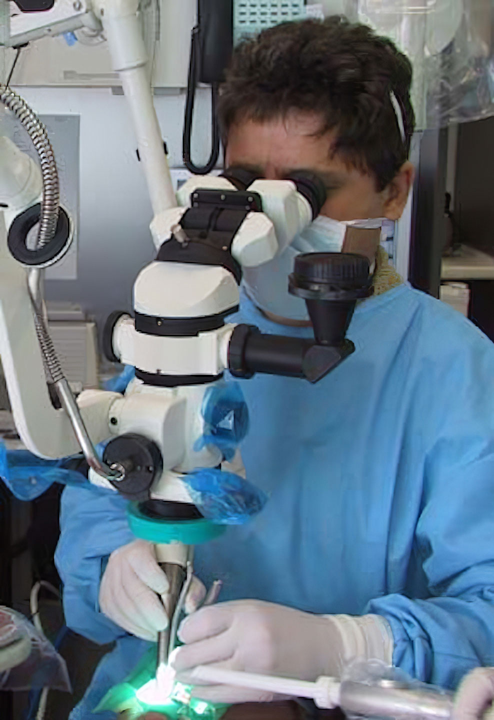
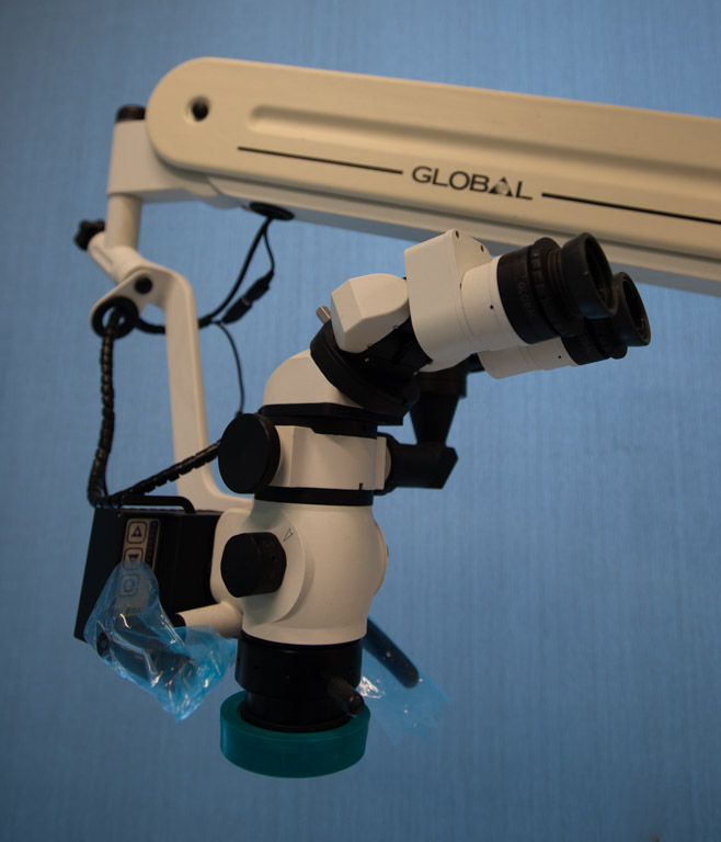
Whenever possible, treatment is performed under magnification using our microscope.
The Global’s six steps of magnification and excellent light source enables us to perform procedures with a far higher degree of accuracy. The microscope also enables true microdentistry, thereby preserving as much of your healthy tooth structure as possible.
A camera attached to the microscope is connected to the overhead TV. If you want to, you can watch on the TV exactly the same image that the dentist is seeing through the eyepieces of the microscope. We can also take still photographs with the camera for use as part of your dental records.
CEREC®
Single-visit Crowns and Fillings
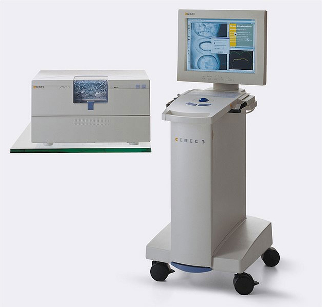
CEREC® CAD/CAM technology is a revolutionary new way to create crowns and fillings in ceramic materials, in a single visit.
Ceramics are stronger and, in many cases, more aesthetic than conventional tooth-coloured fillings
To learn more about our CEREC® CAD/CAM system, click here.
Laser Decay Diagnosis (Diagnodent)
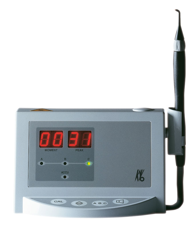
Our Diagnodent unit enables us to detect cavities very early in their development. Being able to detect cavities early, while they are still small, helps a great deal in conserving the precious enamel and dentine.
The fine tip of the unit directs laser light into the tooth and the unit measures changes in the way the light is reflected. These changes are processed in the unit and a digital readout indicates the degree of decay in the tooth being examined.
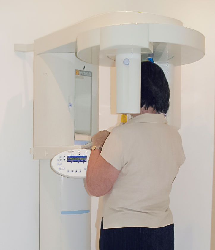
OPG (orthpantamogram) machine
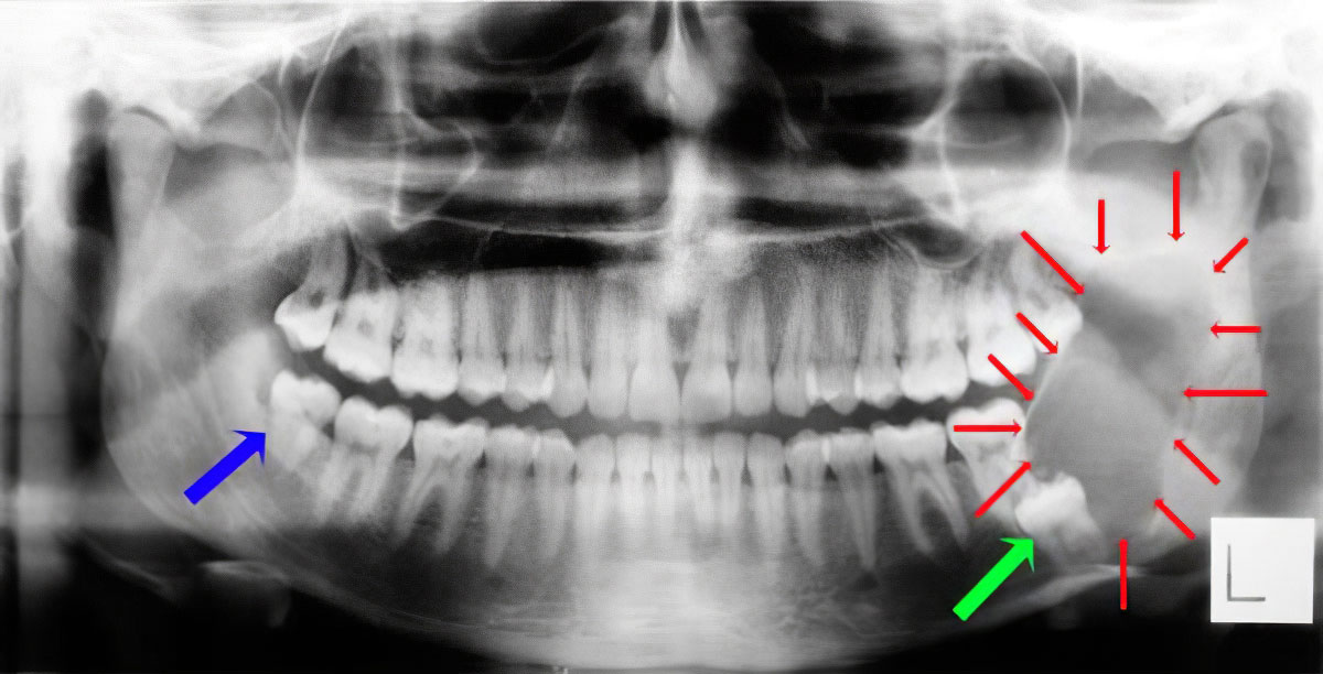
The patient whose OPG is shown above presented complaining of pain in the area of his impacted lower right wisdom tooth (blue arrow). We took an OPG as part of our diagnosis process.
The OPG showed that the lower right wisdom tooth was a problem that could easily be treated, but it revealed a far more serious problem on the left.
An extremely large cyst (outlined by the red arrows) had grown in association with the impacted lower left wisdom tooth (green arrow). The cyst has caused severe destruction of the the bone in his jaw, requiring a very large operation to treat.
Digital Radiography (X-Rays)
Radiographs (X-Rays) are fundamental and essential for the proper and careful diagnosis of our patients’ dental and oral health. Dental radiography, using regular film-based techniques, has been extremely safe. Our modern digital X-Ray facilities have made it even safer. Instead of a film-packet being placed in the mouth, a sensor, very similar in principle to the one found in digital cameras, is used. The sensors are significantly more sensitive than film, which means that significantly shorter exposures are required to produce the image.
Within a second or two after pressing the button, the X-Ray image appears on the computer monitor.
This image can be manipulated to enhance the diagnostic benefit from it. It also allows us to demonstrate with ease to our patients in greater detail the condition of their teeth and the surrounding bone.
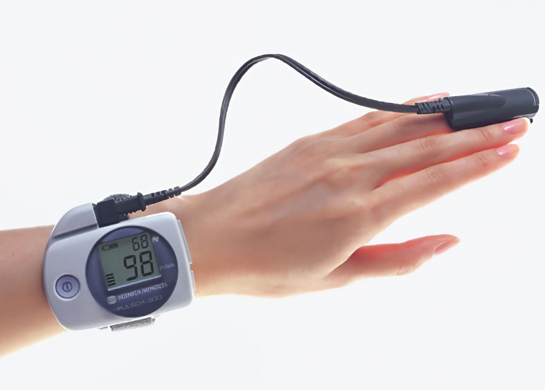
Pulse-Oximeter for sleep survey
We have a Konica-Minolta Pulsox-300i pulse-oPulse-oximeterximeter that we lend to patients for initial sleep screening.
In some cases we can be reasonably certain that the patient has sleep problems that require referral to a sleep physician. There are however, patients whom we suspect might have sleep problems but for whom we would like more information to help determine if a referral to a sleep physician is warranted.
The pulse-oximeter is worn by that patient while they are sleeping. It records the heart rate and the concentration of Oxygen in the blood. In patients with sleep-disordered breathing, it is common for the heart rate to increase and the blood levels of Oxygen to drop when they are going through periods of inadequate airflow.
The recordings from the pulse-oximeter are uploaded to a facility in the USA, who generate a detailed analysis of the information. We don’t charge for the loan of the pulse-oximeter but there is a small fee for having the data analysed in the USA.


